PERIODONTICS
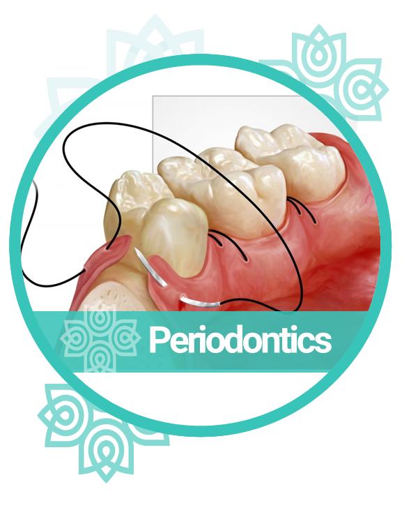
Periodontics is a branch of dentistry that addresses the prevention, diagnosis, and treatment of conditions that affect the tissues supporting your teeth. These tissues include the gums (gingiva), the bone that surrounds your teeth, and the periodontal ligament that connects your teeth to the bone. A healthy foundation is essential for maintaining your smile’s aesthetics and functionality.
| Non-Surgical Treatments – Scaling and Root Planing | |
|---|---|
| Pocket Reduction Procedures | |
| Bone Grafting | |
| Soft Tissue Grafting | |
| Dental Crown Lengthening | |
| Laser Gum Surgery – Gingivectomy |
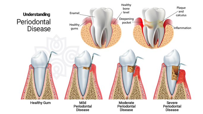
STAGES OF PERIODONTAL DISEASE
| Gingivitis is the mildest form of periodontal disease. It causes the gums to become red, swollen, and bleed easily. There is usually little or no discomfort at this stage. Gingivitis is often caused by inadequate oral hygiene. Gingivitis is reversible with professional treatment and good oral home care. Untreated gingivitis can advance to periodontitis. With time, plaque can spread and grow below the gum line. Toxins produced by the bacteria in plaque irritate the gums. The toxins stimulate a chronic inflammatory response in which the body in essence turns on itself, and the tissues and bone that support the teeth are broken down and destroyed. Gums separate from the teeth, forming pockets (spaces between the teeth and gums) that become infected |
|---|
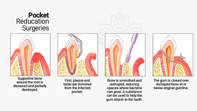
STEPS TO POCKET REDUCTION SURGERY
- Pocket reduction surgery (also known as gingivectomy, osseous surgery and flap surgery) is a collective term for a series of several different surgeries aimed at gaining access to the root of the teeth in order to remove bacteria and tartar (calculus). The human mouth contains dozens of different bacteria at any given time. The bacteria found in plaque (the sticky substance on teeth) produce acids that lead to demineralization of the tooth surface, and ultimately contribute to periodontal disease. Reasons for Pocket Reduction Surgery:
- Reducing bacterial spread
- Halting bone loss
- Facilitate home care
- Enhancing the smile
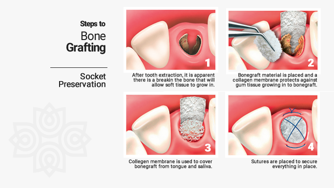
STEPS TO BONE GRAFTING
|
|---|
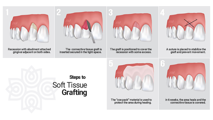
SOFT TISSUE GRAFTING
-
-
-
- Soft tissue grafting is often necessary to combat gum recession. Periodontal disease, trauma, aging, over brushing, and poor tooth positioning are the leading causes of gum recession which can lead to tooth-root exposure in severe cases.When the roots of the teeth become exposed, eating hot and cold foods can be uncomfortable, decay is more prevalent and the aesthetic appearance of the smile is altered. The main goal of soft tissue grafting is to either cover the exposed root or to thicken the existing gum tissue in order to halt further tissue loss. Benefits associated with soft tissue grafting treatment:
-
-
-
-
- Increased comfort
- Improved aesthetics
- Improved gum health
-
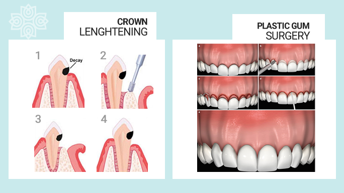
CROWN LENGHTENING & PLASTIC GUM SURGERY
The surgeon removes gum tissue, bone or both to expose more of a tooth. Crown lengthening is done when a tooth needs to be fixed. Sometimes, not enough of the tooth sticks out above the gum to support a filling or crown. There are two main reasons a patient may need to undergo the crown lengthening procedure:
| The first reason is to make it possible to place a restoration, such as a crown, if there isn’t enough exposed tooth structure to support it. By removing some of the gum and/or bone near the tooth, we can create enough space to successfully complete the restoration. | |
|---|---|
| The second reason is cosmetic in nature. Patients who have what is known as a “gummy smile,” or a smile in which the gums cover too much of the teeth and make the teeth appear too short, can greatly benefit from crown lengthening. We can remove some of the gum tissue to make the gum line even and show more of the teeth, which makes the patient’s smile appear larger and more attractive. |
LASER GUM SURGERY
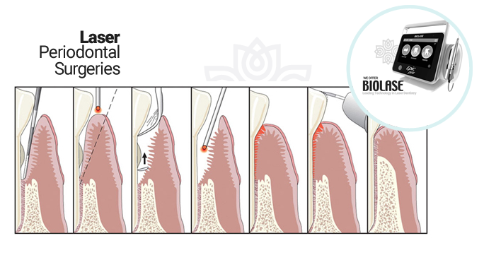
In periodontal laser therapy, the provider uses a dental laser to access and remove the inflamed gum tissue from around the root of the tooth. When the infected tissue is removed and the root is exposed, the root scaling begins. … The area between the gum and the root can then regenerate during the healing process.
According to the Academy of General Dentistry (AGD), there are ample benefits to using lasers for excising diseased gum tissue.
- No general anesthetic is needed.
- No general anesthetic is needed.
- Lasers can target the diseased areas precisely and accurately.
- Bleeding, pain & swelling are limited since PLT is less invasive.
- Recovery and healing times are shorter.
SINUS LIFT SURGERY
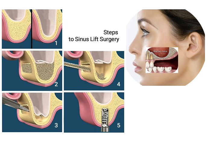
A sinus lift, also known as a sinus augmentation or sinus floor elevation, is a surgical dental procedure used to increase the amount of bone in the upper jaw (maxilla) in the area of the molars and premolars. This procedure is typically performed when there is insufficient bone height in the upper jaw to support the placement of dental implants.
Sinus lifts are crucial for individuals with insufficient bone in the upper jaw, particularly in the posterior region, to enable them to receive dental implants. This procedure has a high success rate, and it has significantly improved the ability to replace missing teeth in the upper jaw, even in cases where bone loss has occurred due to tooth extraction or other factors.
Here's an overview of the sinus lift procedure:
Before the procedure, a thorough examination, including dental X-rays or CT scans, is performed to evaluate the patient’s bone structure and sinus anatomy. This helps the oral surgeon determine if a sinus lift is necessary and plan the surgery accordingly.
The patient is administered local anesthesia to numb the area where the procedure will be performed, ensuring they do not feel pain during the surgery.
A small incision is made in the gum tissue near the area where the dental implants will be placed. This creates a flap that provides access to the underlying bone.
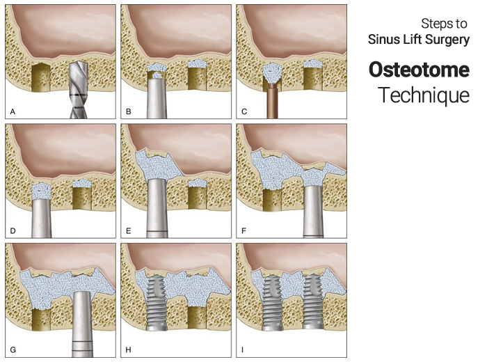
The oral surgeon carefully lifts the membrane lining the sinus (the Schneiderian membrane) away from the underlying bone. This creates a small space between the membrane and the bone.
With the sinus membrane gently lifted, a bone graft material is placed into the space created. The bone graft can be taken from the patient’s own body (autograft), from a donor (allograft), or it can be a synthetic material (alloplastic graft). The choice of graft material depends on various factors, including the patient’s needs and surgeon’s preference.
Closing the Incision: After the bone graft is placed, the incision is sutured closed. The bone graft material will eventually integrate with the patient’s existing bone over several months, creating a more stable and thicker bone structure.
The patient will need some time to heal, typically several months. During this time, the bone graft will fuse with the patient’s natural bone, creating a solid foundation for the future dental implants.
Dental Implant Placement: Once the bone has healed and is deemed strong enough, the patient can undergo the placement of dental implants into the augmented area. These implants can eventually support crowns, bridges, or dentures.
